Peptide Vial Math is a set of free, accessible calculators for one job: turning "mg in a vial" and "mL of water" into exact concentrations, volumes and syringe readings.
Reconstitution math is simple, but errors are easy to make and costly: mg vs mcg is a 1,000× error, units vs mL a 100× one. FDA has documented people drawing 50 units instead of 5. We wanted a tool that shows every intermediate number, warns when a reading can't be measured cleanly, and states its answer in one plain sentence you can check by hand.
Peptide Vial Math Editorial Team
Every page on Peptide Vial Math is written and reviewed by the Peptide Vial Math Editorial Team, and is credited to the team rather than to individuals. We don't attach personal names or professional credentials to pages, and we don't present the site as medical advice. What we can show you instead is the work: every factual claim about a compound, a diluent or a regulation links to a primary source (FDA, DailyMed, CDC, ClinicalTrials.gov, PubMed or PubChem), and every number comes from one tested code module.
How a page is reviewed, step by step, is documented on the methodology page. Each page shows the date it was last reviewed.
What we'll never publish
Dosing recommendations, schedules or "protocols".
Seller rankings, scores, star ratings or buy links.
Testimonials, invented experts or unverifiable statistics.
Picture credits
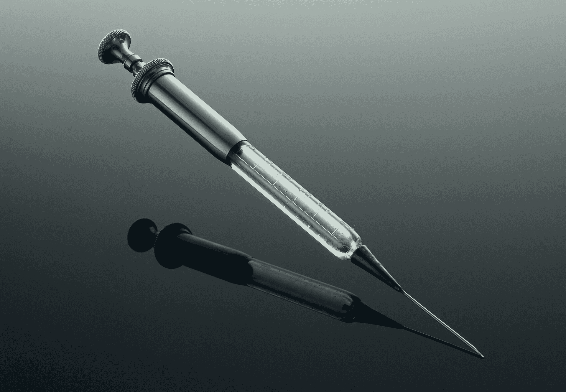
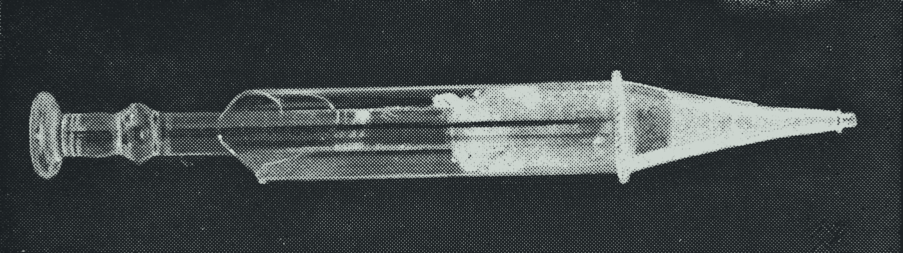
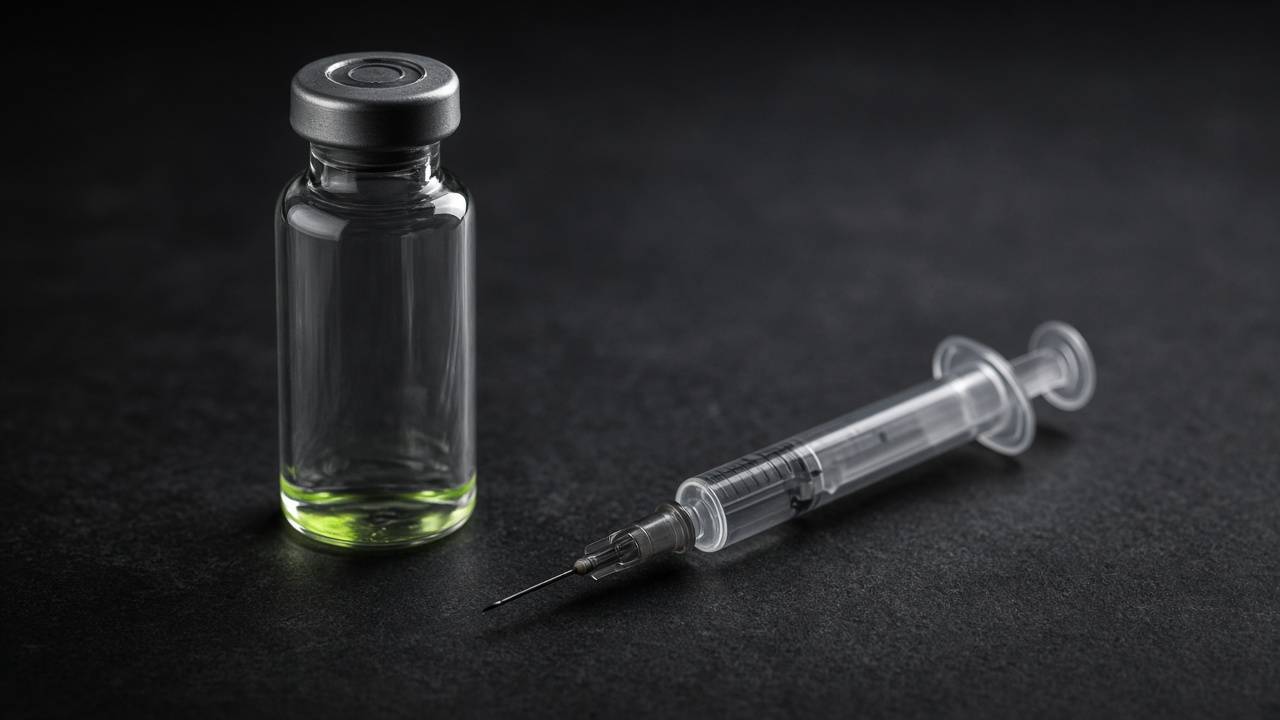
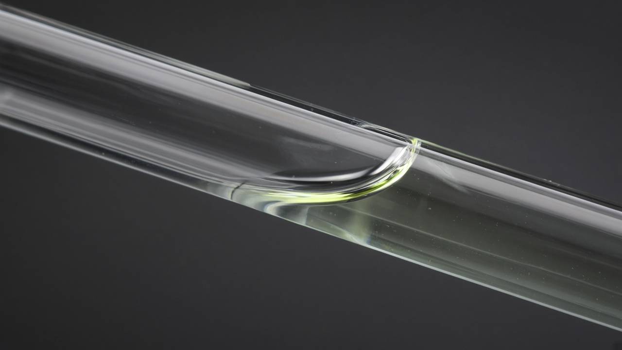
The pictures on Peptide Vial Math are historical laboratory plates and museum photographs of graduated glassware and early syringes, chosen because they show the same problem the calculators solve: reading a volume off a scale. They are not our products and show no product sold today. Each one is converted to the site's graphite tone and cropped to remove printed figure labels. The modern photographs (unlabeled vials, a syringe, a meniscus) are generated images made for Peptide Vial Math: they show no real product, brand, label or person.
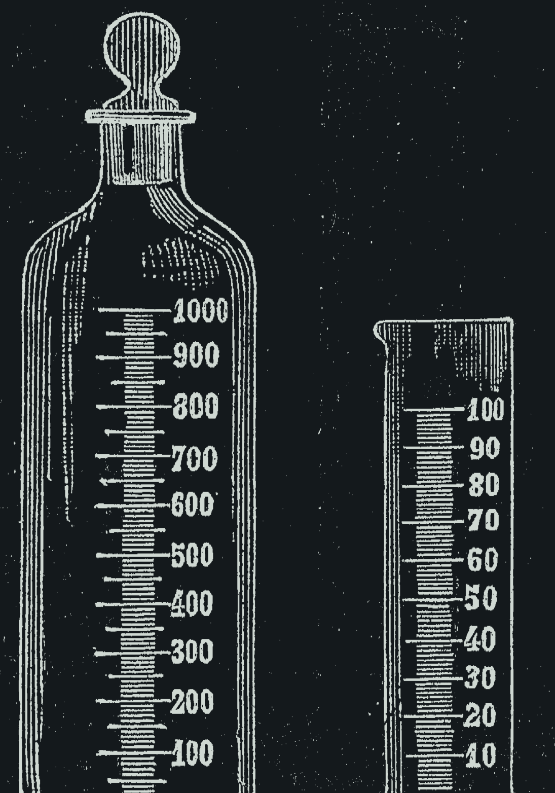
Graduated chemical glassware, Alessandri 1895, fig. 1. Public domain.Alessandri (1895), scan by valeg96 via Wikimedia Commons.
Burettes, Alessandri 1895, figs. 12 and 13. Public domain.Alessandri (1895), scan by valeg96 via Wikimedia Commons.
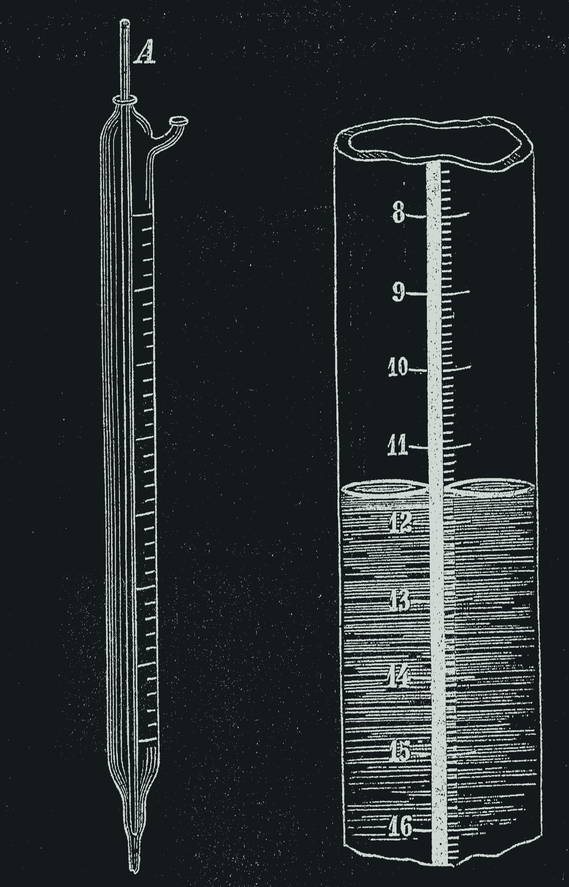
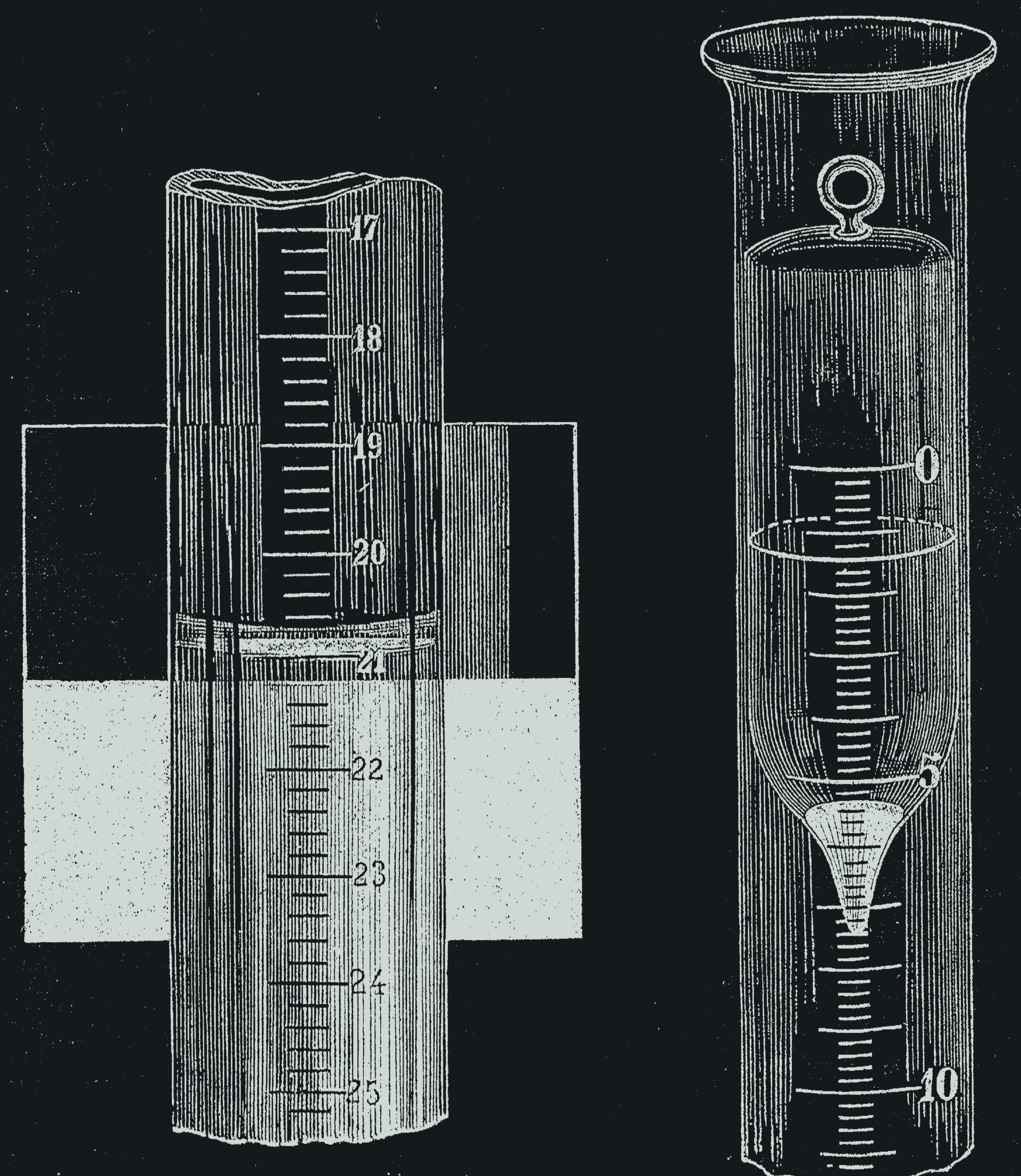
Reading the meniscus, Alessandri 1895, figs. 14 and 15. Public domain.Alessandri (1895), scan by valeg96 via Wikimedia Commons.
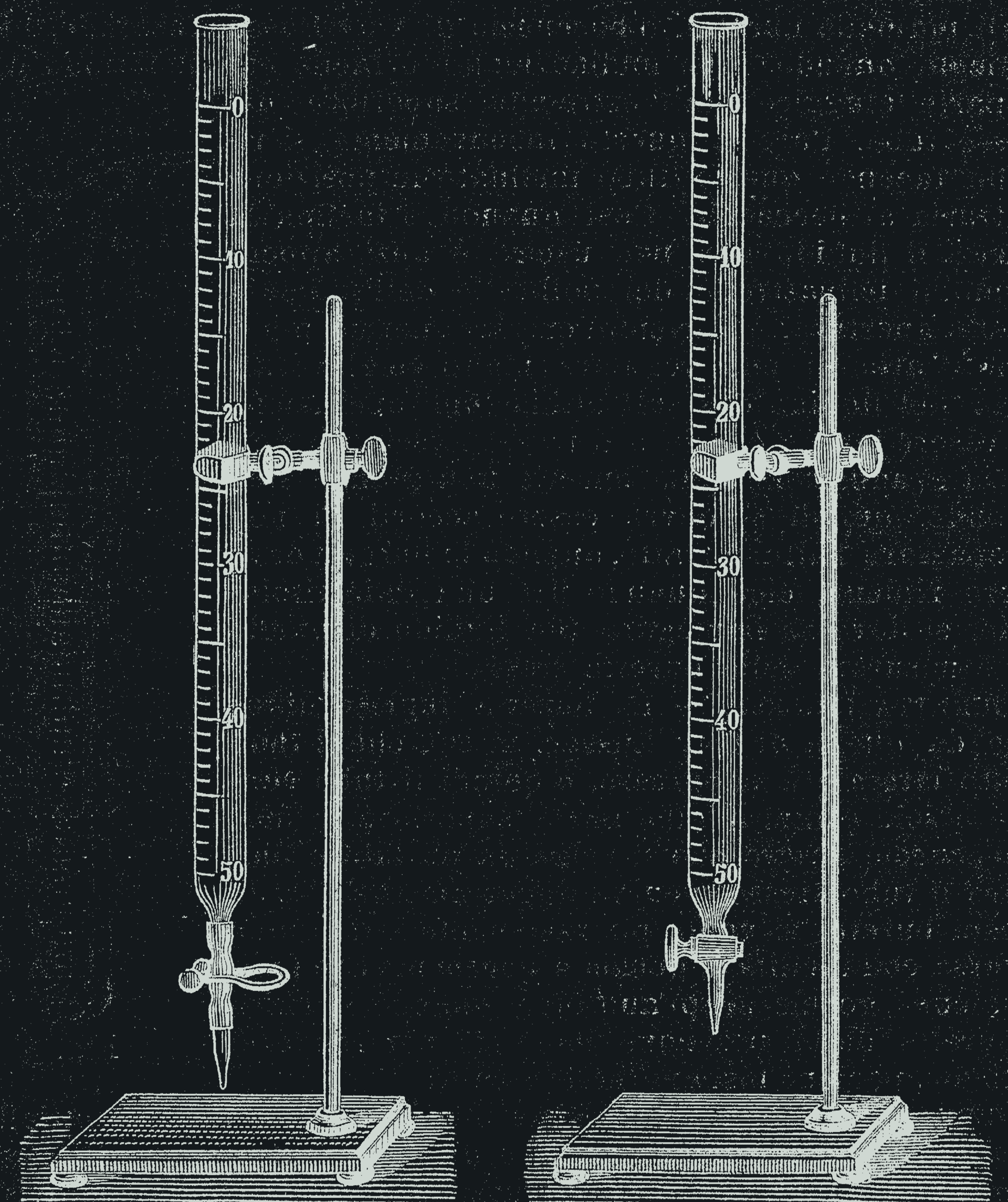
Mohr burette, Alessandri 1895, figs. 9 and 10. Public domain.Alessandri (1895), scan by valeg96 via Wikimedia Commons.
Hypodermic syringe, England, 1855–1865. CC BY 4.0.Wellcome Collection (L0057801) via Wikimedia Commons.
Hypodermic syringe of Dr Alexander Wood (Wellcome M0009706). CC BY 4.0.Wellcome Collection (M0009706) via Wikimedia Commons.
Unlabeled vial and syringe. Generated image made for Peptide Vial Math.Generated image made for Peptide Vial Math.
A meniscus, close up. Generated image made for Peptide Vial Math.Generated image made for Peptide Vial Math.
Powder, solution, empty vial. Generated image made for Peptide Vial Math.Generated image made for Peptide Vial Math.
A vial in cold storage. Generated image made for Peptide Vial Math.Generated image made for Peptide Vial Math.
Adding diluent to a freeze-dried powder. Generated image made for Peptide Vial Math.Generated image made for Peptide Vial Math.
Corrections
If you find an error in a formula, a table or a fact, email corrections@peptidevialmath.com. We fix confirmed errors promptly and update the page's "updated" date.